


-
Academic Article
User Achievement | Exploring Plants' "Invisible Coat": Coherent Raman Microscopy Opens a New Era for Plant Cell Wall Research

Research Background and Core Conclusion
The article presents a plant cell wall study published in Science China Life Sciences. The team led by Jinxing Lin at Beijing Forestry University used Raman microscopy to analyze the major components of plant cell walls and achieved in situ, single-cell characterization of cellulose, hemicellulose, pectin, lignin, and related components. The page emphasizes that Vibronix's coherent Raman imaging technology played an important supporting role in the work.
Figure 1: Research outcome, sourced from Science China Life Sciences.
Core Conclusion: Coherent Raman provides a new in situ, label-free, single-cell-scale route for plant cell wall analysis.
Technical Principle and Method Comparison
The article explains that plant cell walls are composed of cellulose, hemicellulose, pectin, lignin, and other structural components, and then compares confocal Raman microscopy (CRM) with coherent Raman scattering (SRS). Because coherent Raman imaging does not require staining and uses femtosecond dual-pulse excitation to resonantly amplify Raman signals by roughly one million times, it greatly improves imaging speed while reducing interference from plant autofluorescence.
1. What Is the Plant Cell Wall: The external support structure of plant cells that maintains morphology and protects intracellular contents.
2. CRM Limitation: Autofluorescence interference is strong, and imaging can take tens of minutes to hours.
3. SRS Advantage: Label-free imaging with higher speed, well suited to large-area samples and dynamic processes.
Imaging Results and Research Significance
The page includes Raman and coherent Raman images of plant cell wall components in pine xylem and Jatropha seed coats. It states that SRS and CRM produce highly consistent results when identifying deposition of different lignin units, but SRS reduces single-point integration time to about 10 us and completes full-image acquisition in roughly 1 second. The article concludes that coherent Raman microscopy offers a powerful new tool for plant science, biophysics, and materials research.
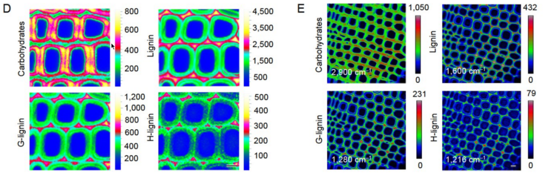
Figure 2: Confocal Raman and coherent Raman imaging of component distribution in pine xylem cell walls
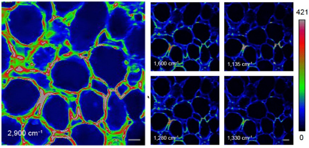
Figure 3: Coherent Raman imaging and rainbow analysis of plant cell wall components in Jatropha seed coats
Significance: Supports discrimination of sugars, cellulose, and different lignin types, helping clarify structure-function relationships in plant cell walls.
Popular Tags
-
Coherent Raman
-
Botany
Recommendation
-
-
Academic Article
2026-02-09User Achievement | The Guizhou University Team Uses Stimulated Raman Imaging to Decipher the Efficiency-Boosting Mechanism of Enzyme Catalysis -
-
-
Academic Article
2026-01-22User Achievement | A New Breakthrough from the Peking University Team: Coherent Raman Reveals the Molecular Basis of Light-Regulated Hypocotyl Elongation -