


Synthetic Biology
Resolve metabolic heterogeneity in engineered microbes at single-cell resolution, track intracellular metabolic flux and the spatial distribution of key enzymes in real time, and accelerate pathway optimization and high-yield strain development for bio-based manufacturing.

In biomedical research, preserving sample authenticity, observing deep structures, tracking molecular dynamics, and achieving quantitative accuracy remain fundamental requirements. Conventional imaging methods are often constrained by fluorescent-label interference, slow acquisition speeds, and limited penetration depth, which in turn restrict breakthroughs in disease-mechanism studies, drug development, and clinical diagnostics. Coherent Raman imaging addresses these challenges through label-free contrast, avoiding phototoxicity and signal interference from fluorescent labeling while preserving the physiological activity of samples such as organoids and living tissues. With submicron spatial resolution and millimeter-scale penetration depth, it can clearly reveal fine structures including tumor infiltration boundaries and neuronal layering, while also distinguishing the distributions of proteins, lipids, and nucleic acids in deeper tissue regions. By recognizing molecular vibrational signatures with high specificity, the technology supports real-time tracking of dynamic processes such as glucose metabolism and lipid synthesis, enabling precise quantification of endogenous molecules. From stem-cell differentiation studies in organoid development and label-free detection of amyloid plaques in Alzheimer's disease to drug-penetration assessment in 3D models and analysis of tumor heterogeneity, this approach provides a new research paradigm for disease-model construction, drug-screening optimization, and precision clinical pathology, helping biomedical research move from static analysis to dynamic tracking and from broad screening to precise quantification.
Solution Advantages


-
 Label-Free Imaging
Avoid fluorescence-induced phototoxicity and photobleaching while preserving the physiological authenticity of the sample.

-
 Deep Penetration with High Resolution
Combine tissue-clearing workflows with millimeter-scale imaging depth and submicron spatial resolution.
-
 Precise Quantification
Signal intensity remains linearly correlated with concentration, enabling direct quantification of endogenous molecules such as proteins, lipids, and metabolites.
-
 Ultrafast Imaging
Signal enhancement of up to 106 supports imaging in seconds to minutes rather than the hours often required by conventional approaches.


Typical Scenarios
-

 Glycolipid Metabolism
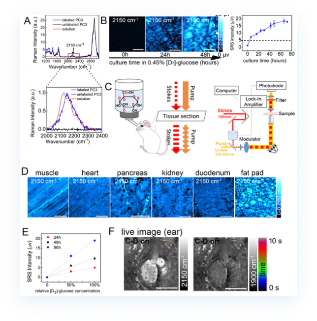
For metabolic processes involving small biomolecules, coherent Raman imaging can track the metabolic trajectory of deuterated glucose in cells and visualize molecular distribution changes at different time points in real time.
-
 Pathology Research
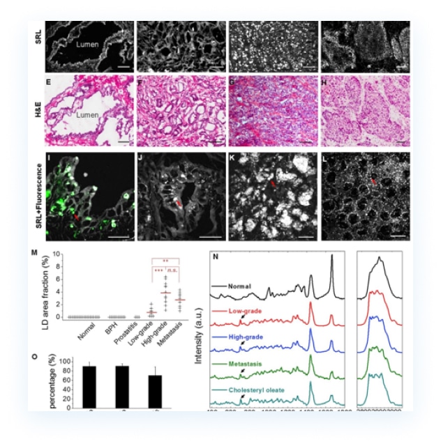
By comparing the spectral differences between diseased and normal tissues, the system can help identify tumor biomarkers. Quantitative analysis can also correlate with tumor malignancy and support further signaling-pathway research.
-
 Glycolipid Metabolism
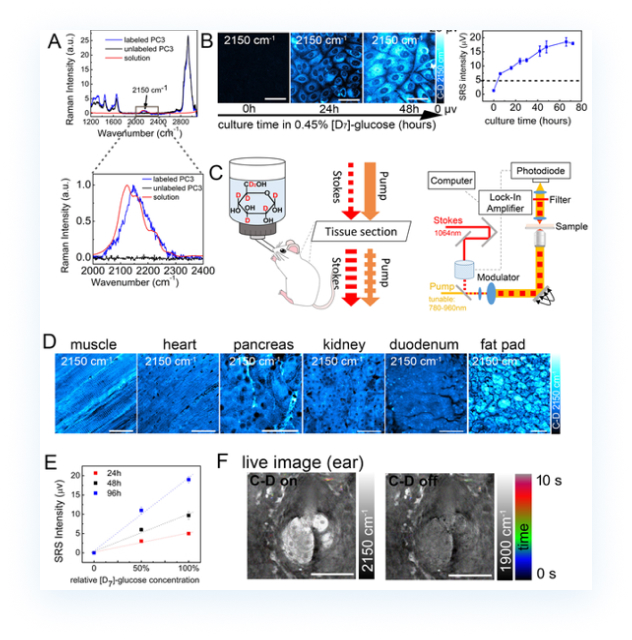
For metabolic processes involving small biomolecules, coherent Raman imaging can track the metabolic trajectory of deuterated glucose in cells and visualize molecular distribution changes at different time points in real time.