



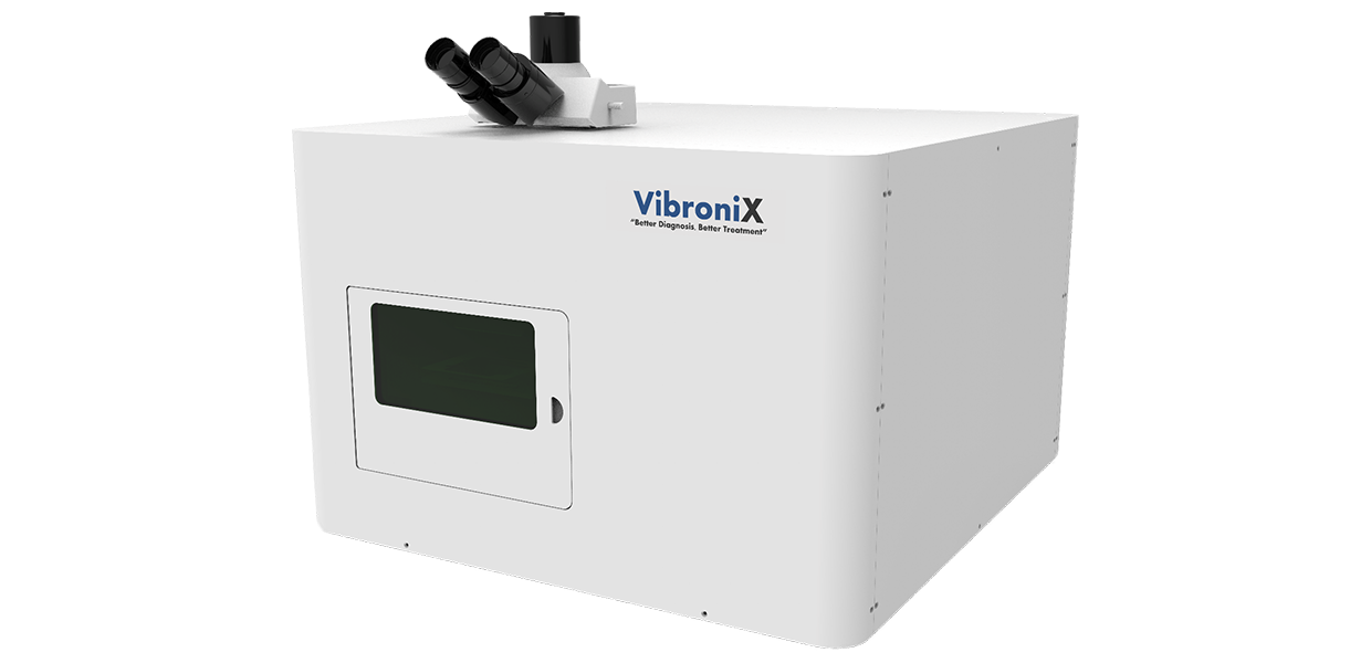
LMD-MK-Ⅰ
LMD-MK-I is an advanced research platform that integrates high-precision laser cutting, intelligent imaging, and automated sample collection. Through precise laser microdissection, it can isolate target tissues, cells, or subcellular structures from complex samples. Combined with high- resolution imaging and intelligent operation, it provides reliable sample-isolation workflows for molecular biology, medical research, plant science, and related fields.


Principle of Operation
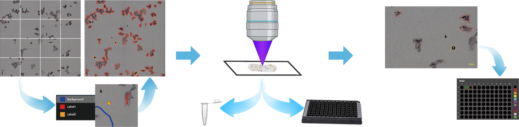
Traditional workflows require at least six steps and repeated switching among the main program, image-analysis software, and scripts. Coordinate calibration and file import alone can take more than 30 minutes. The LMD smart imaging-guided laser microdissection system reduces the workflow to three steps: open the main program, select the target region, and cut automatically. No software switching is required, and the process can be completed within 15 minutes, improving efficiency by 200%.
Core Advantages and Customer Value


-
 High-Precision Laser Cutting
An innovative galvanometer-control architecture enables real-time laser-beam control with micron-scale path positioning while minimizing thermal spread to surrounding tissue.
The page claims edge smoothness improvements above 30%, helping preserve sample integrity in both hard tissues such as calcified nodules and bone sections and softer structures such as nerve fibers.
-
 Intelligent Imaging and Automation
Autofocus adjusts focal position automatically according to sample thickness to maintain a clear full-field view.
Full-slide scanning and automatic stitching help reduce error, while AI-based tissue-texture and cell-shape recognition can generate cutting paths automatically.
-
 High-Throughput Smart Collection
Programmable 96-well plate collection enables contact-free, contamination-controlled transfer, automatically assigning samples to target wells based on color-coded labels and recording position mappings for traceability.
Applications
-

 Pharma and Life Science
Cancer research: isolate specific tumor-cell populations for microenvironment and genotyping studies.
Neuroscience: analyze neuronal structure and signaling mechanisms.
Molecular biology: prepare high-fidelity samples for nucleic-acid and protein analysis. -
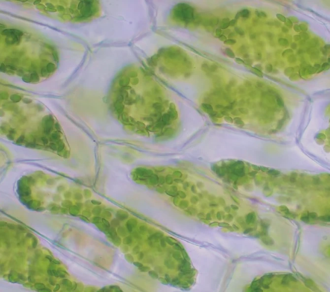 Plant Science
Study cell and tissue structures in plants and the distribution of their components.
-
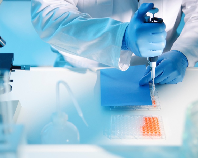 Forensics and Cell Culture
Identify biomarkers in target tissues for case investigation and support cell cloning, passaging, and downstream experimental workflows.
-
Pharma and Life Science
Cancer research: isolate specific tumor-cell populations for microenvironment and genotyping studies.
Neuroscience: analyze neuronal structure and signaling mechanisms.
Molecular biology: prepare high-fidelity samples for nucleic-acid and protein analysis. -
Plant Science
Study cell and tissue structures in plants and the distribution of their components.
-
Forensics and Cell Culture
Identify biomarkers in target tissues for case investigation and support cell cloning, passaging, and downstream experimental workflows.
Technical Specifications

| Laser Configuration | Cutting Mode | Electronic Eyepiece | Stage | Objectives | Microscope | Main Functions | ||
| Option 1 Wavelength: 355 nm pulse energy: 100 μJ repetition rate: 1 ~ 100 Hz pulse width: <4 ns laser power adjustable from 0% to 100% | Option 2 Wavelength: 355 nm pulse energy: 120 μJ repetition rate: 1 ~ 5000 Hz pulse width: <4 ns laser power adjustable from 0% to 100% | The galvanometer controls the laser beam for arbitrary path outlining and scanning in the X-Y plane | Color CMOS camera, 30 fps, >=2K resolutio | Sample Stage 2 × slide travel: 100 mm × 100 mm × 20 mm XY resolution: 0.05 μm Z resolution: 0.1 μm XY repeat positioning accuracy ≤ 3 μm Z repeat positioning accuracy ≤ 1 μm | Collection Stage 1 × microplate travel: 110 mm ×120mm ×20 mm XY resolution: 0.1μm Z resolution: 0.02 μm XY repeat positioning accuracy ≤0.5μm Z repeat positioning accuracy ≤0.4μm | Standard 4X and 10X air objectives with motorized switching, optional upgrades available | Upright research-grade microscope with transmitted illumination and optional fluorescence module | Standard-shape and freeform cutting, adjustable cut-line width / laser power / cutting speed, autofocus and auto-exposure, large-area mosaic imaging, automatic recognition and cutting of target regions, and programmable sample collection |
Downloads
-

Product Brochure
LMD Smart Imaging Laser Microdissection SystemProduct:LMD-MK-Ⅰ